Product overview

Full product name
Code
Host
Isotype
Size
Clonality
Immunogen
Purification
Concentration
Formulation
Storage
Background
This gene encodes a member of the epidermal growth factor (EGF) receptor family of receptor tyrosine kinases. This protein has no ligand binding domain of its own and therefore cannot bind growth factors. However, it does bind tightly to other ligand-bound EGF receptor family members to form a heterodimer, stabilizing ligand binding and enhancing kinase-mediated activation of downstream signalling pathways, such as those involving mitogen-activated protein kinase and phosphatidylinositol-3 kinase. Allelic variations at amino acid positions 654 and 655 of isoform a (positions 624 and 625 of isoform b) have been reported, with the most common allele, Ile654/Ile655, shown here. Amplification and/or overexpression of this gene has been reported in numerous cancers, including breast and ovarian tumors. Alternative splicing results in several additional transcript variants, some encoding d
Uniprot accession
Molecular weight
Gene ID
Synonyms
Research area
Target protein
Recommended dilution
| Application | Dilution |
|---|---|
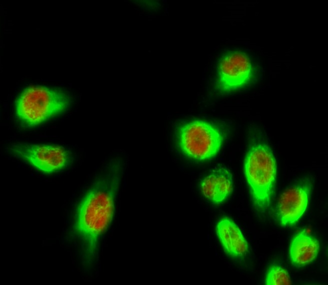
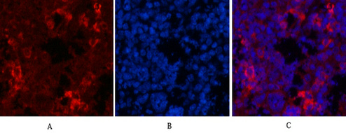
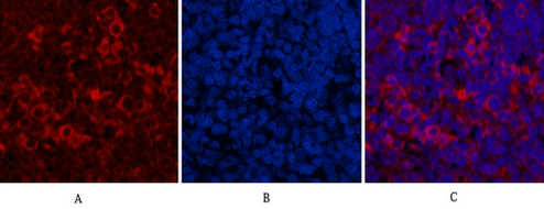
| IF | 1:200 |
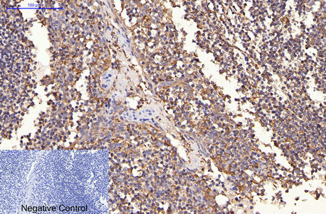
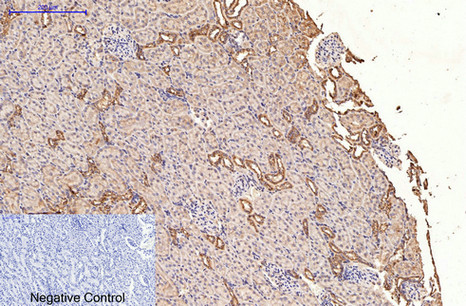
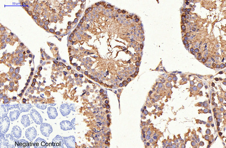
| IHC | 1:200 |
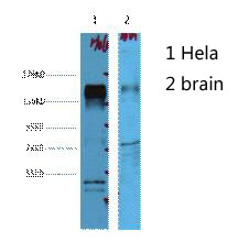
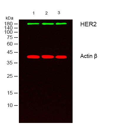
| WB | 1:2000-4000 |
Images
Protocols
Customer reviews and Q&As