Product overview

Full product name
Code
Host
Isotype
Size
Clonality
Immunogen
Purification
Concentration
Formulation
Storage
Background
Histones are basic nuclear proteins that are responsible for the nucleosome structure of the chromosomal fiber in eukaryotes. This structure consists of approximately 146 bp of DNA wrapped around a nucleosome, an octamer composed of pairs of each of the four core histones (H2A, H2B, H3, and H4). The chromatin fiber is further compacted through the interaction of a linker histone, H1, with the DNA between the nucleosomes to form higher order chromatin structures. This gene is intronless and encodes a replication-dependent histone that is a member of the histone H3 family. Transcripts from this gene lack polyA tails| instead, they contain a palindromic termination element. This gene is found in the large histone gene cluster on chromosome 6p22-p21.3.
Uniprot accession
Molecular weight
Gene ID
Synonyms
Research area
Target protein
Recommended dilution
| Application | Dilution |
|---|---|
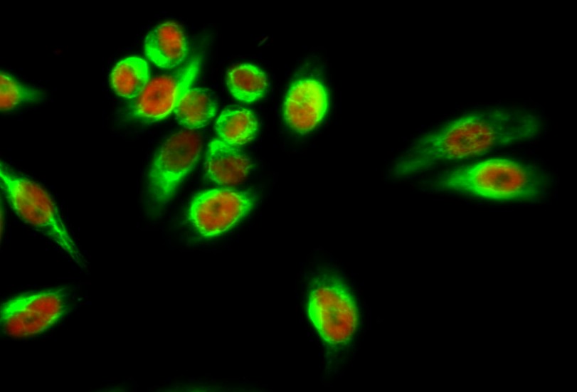
| IF | 1:100-500 |
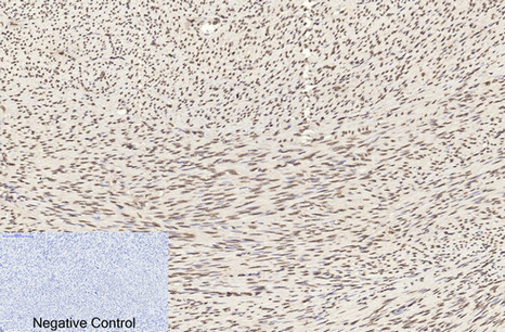
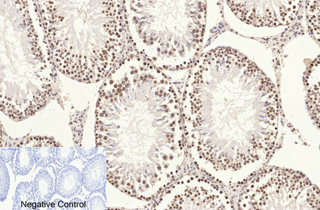
| IHC | 1:50-300 |
| IP | 1:200 |
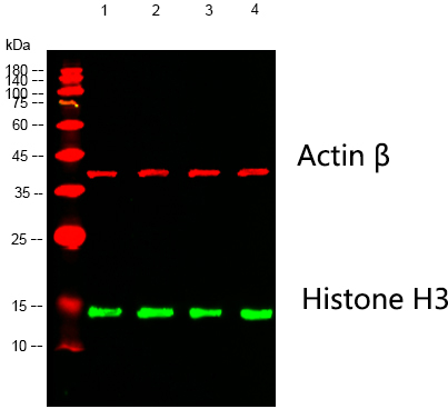
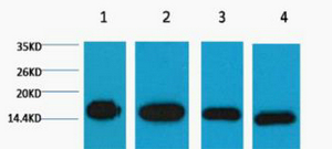
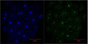
| WB | 1:2000-5000 |
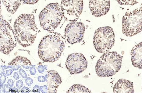
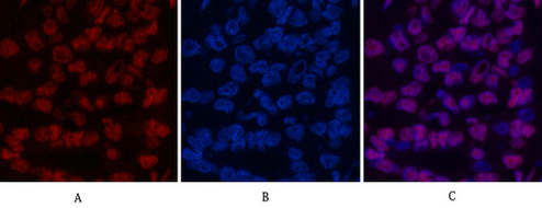
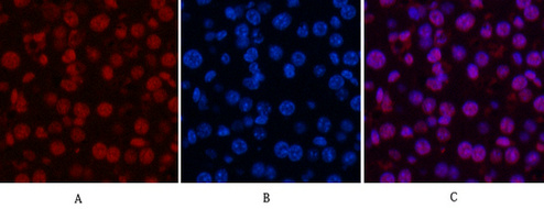
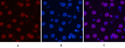
Images
Protocols
Customer reviews and Q&As