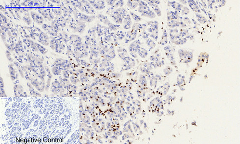
Immunohistochemical analysis of paraffin-embedded Human-stomach-cancer tissue. 1,Histone H2A.X (phospho Ser139) Polyclonal Antibody was diluted at 1:200(4°C overnight). 2, Sodium citrate pH 6.0 was used for antibody retrieval(>98°C,20min). 3,Secondary antibody was diluted at 1:200(room temperature, 30min). Negative control was used by secondary antibody only.
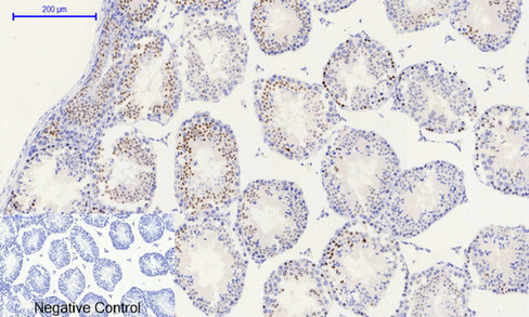
Immunohistochemical analysis of paraffin-embedded Rat-testis tissue. 1,Histone H2A.X (phospho Ser139) Polyclonal Antibody was diluted at 1:200(4°C overnight). 2, Sodium citrate pH 6.0 was used for antibody retrieval(>98°C,20min). 3,Secondary antibody was diluted at 1:200(room temperature, 30min). Negative control was used by secondary antibody only.
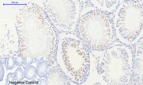
Immunohistochemical analysis of paraffin-embedded Mouse-testis tissue. 1,Histone H2A.X (phospho Ser139) Polyclonal Antibody was diluted at 1:200(4°C overnight). 2, Sodium citrate pH 6.0 was used for antibody retrieval(>98°C,20min). 3,Secondary antibody was diluted at 1:200(room temperature, 30min). Negative control was used by secondary antibody only.
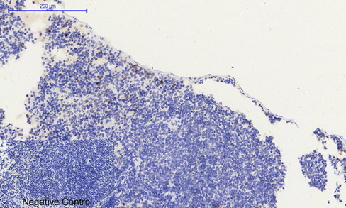
Immunohistochemical analysis of paraffin-embedded Mouse-spleen tissue. 1,Histone H2A.X (phospho Ser139) Polyclonal Antibody was diluted at 1:200(4°C overnight). 2, Sodium citrate pH 6.0 was used for antibody retrieval(>98°C,20min). 3,Secondary antibody was diluted at 1:200(room temperature, 30min). Negative control was used by secondary antibody only.
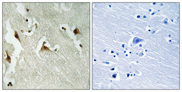
Immunohistochemical analysis of paraffin-embedded Human Amygdala. 1, Antibody was diluted at 1:200(4°C overnight). 2, High-pressure and temperature EDTA, pH8.0 was used for antigen retrieval. 3,Secondary antibody was diluted at 1:200(room temperature, 30min).
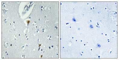
Immunohistochemical analysis of paraffin-embedded Human brain. Antibody was diluted at 1:100(4°C overnight). High-pressure and temperature Tris-EDTA,pH8.0 was used for antigen retrieval. Negetive contrl (right) obtaned from antibody was pre-absorbed by immunogen peptide.
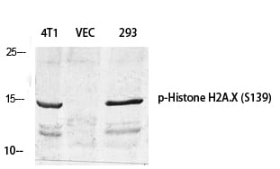
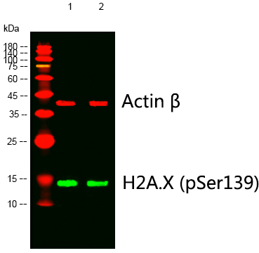
Western blot analysis of lysates from 1) 4T1, 2) 293 cells, (Green) primary antibody was diluted at 1:1000, 4°C overnight, Dylight 800 secondary antibody was diluted at 1:10000, 37°C 1hour. (Red) Actin β Monoclonal Antibody(5B7) was diluted at 1:5000 as loading control, 4°C overnight,Dylight 680 secondary antibody was diluted at 1:10000, 37°C 1hour.
Western Blot analysis of various cells using Phospho-Histone H2A.X (S139) Polyclonal Antibody diluted at 1:500
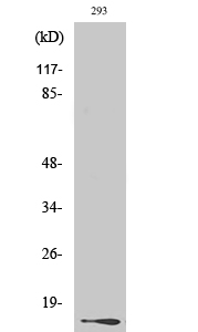
Western Blot analysis of 293 cells using Phospho-Histone H2A.X (S139) Polyclonal Antibody diluted at 1:500
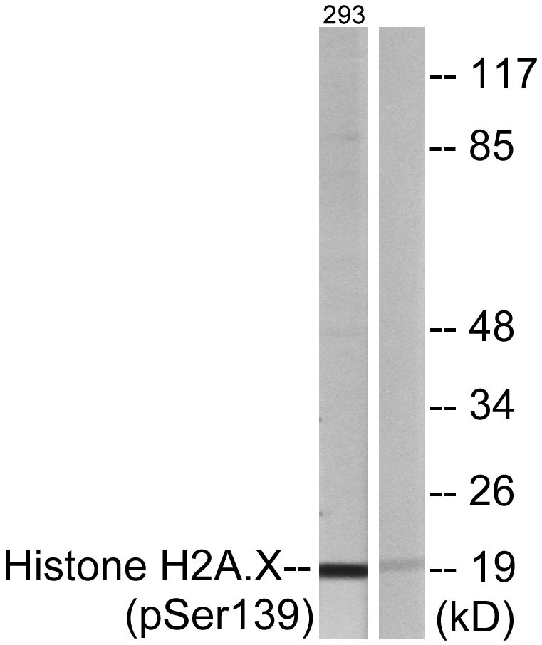
Western blot analysis of lysates from 293 cells treated with heat shock, using Histone H2A.X (Phospho-Ser139) Antibody. The lane on the right is blocked with the phospho peptide.