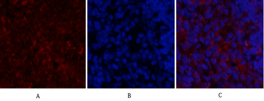
Immunofluorescence analysis of human-lung tissue. 1,SDF-1 Polyclonal Antibody(Red) was diluted at 1:200(4°C overnight). 2, Cy3 labled Secondary antibody was diluted at 1:300(room temperature, 50min).3, Picture B: DAPI(blue) 10min. Picture A:Target. Picture B: DAPI. Picture C: merge of A+B
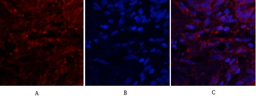
Immunofluorescence analysis of rat-spleen tissue. 1,SDF-1 Polyclonal Antibody(Red) was diluted at 1:200(4°C overnight). 2, Cy3 labled Secondary antibody was diluted at 1:300(room temperature, 50min).3, Picture B: DAPI(blue) 10min. Picture A:Target. Picture B: DAPI. Picture C: merge of A+B
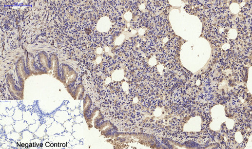
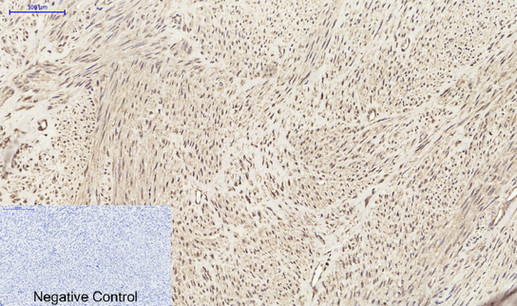
Immunohistochemical analysis of paraffin-embedded Human-uterus tissue. 1,SDF-1 Polyclonal Antibody was diluted at 1:200(4°C overnight). 2, Sodium citrate pH 6.0 was used for antibody retrieval(>98°C,20min). 3,Secondary antibody was diluted at 1:200(room temperature, 30min). Negative control was used by secondary antibody only.
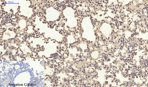
Immunohistochemical analysis of paraffin-embedded Rat-lung tissue. 1,SDF-1 Polyclonal Antibody was diluted at 1:200(4°C overnight). 2, Sodium citrate pH 6.0 was used for antibody retrieval(>98°C,20min). 3,Secondary antibody was diluted at 1:200(room temperature, 30min). Negative control was used by secondary antibody only.
Immunohistochemical analysis of paraffin-embedded Mouse-lung tissue. 1,SDF-1 Polyclonal Antibody was diluted at 1:200(4°C overnight). 2, Sodium citrate pH 6.0 was used for antibody retrieval(>98°C,20min). 3,Secondary antibody was diluted at 1:200(room temperature, 30min). Negative control was used by secondary antibody only.
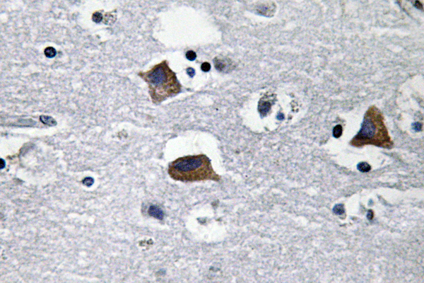
Immunohistochemistry analysis of SDF-1 antibody in paraffin-embedded human brain tissue.
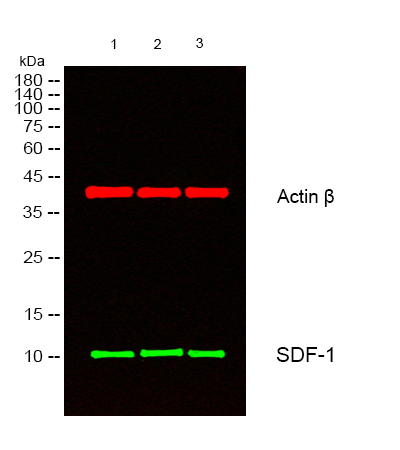
Western blot analysis of lysates from 1) KB, 2) Hela ,3) MCF-7 cells, (Green) primary antibody was diluted at 1:1000, 4°C overnight, secondary antibody was diluted at 1:10000, 37°C 1hour. (Red) Actin β Monoclonal Antibody(5B7) was diluted at 1:5000 as loading control, 4°C overnight,secondary antibody was diluted at 1:10000, 37°C 1hour.
Western Blot analysis of various cells using SDF-1 Polyclonal Antibody diluted at 1:2000
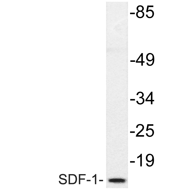
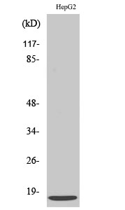
Western Blot analysis of HepG2 cells using SDF-1 Polyclonal Antibody diluted at 1:2000
Western blot analysis of lysate from HepG2 cells, using SDF-1 antibody.
Western blot analysis of lysates from 1) KB, 2) Hela ,3) MCF-7 cells, (Green) primary antibody was diluted at 1:1000, 4°C overnight, secondary antibody was diluted at 1:10000, 37°C 1hour. (Red) Actin β Monoclonal Antibody(5B7) was diluted at 1:5000 as loading control, 4°C overnight,secondary antibody was diluted at 1:10000, 37°C 1hour.