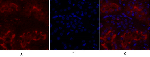
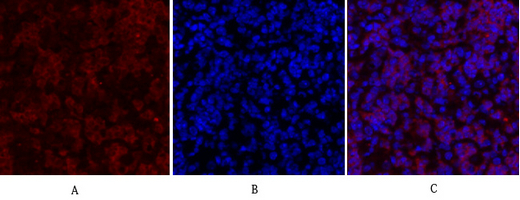
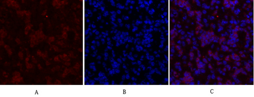
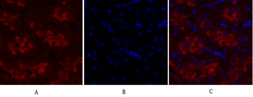
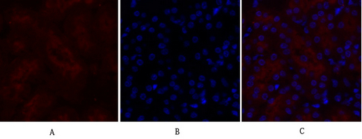
Immunofluorescence analysis of rat-kidney tissue. 1,IGF-IR Polyclonal Antibody(red) was diluted at 1:200(4°C,overnight). 2, Cy3 labled Secondary antibody was diluted at 1:300(room temperature, 50min).3, Picture B: DAPI(blue) 10min. Picture A:Target. Picture B: DAPI. Picture C: merge of A+B
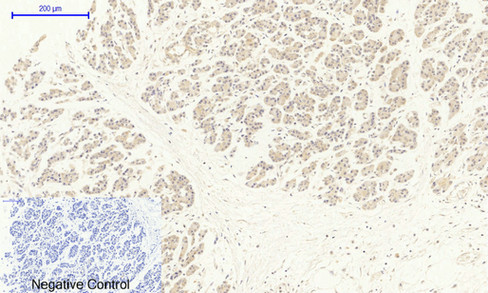
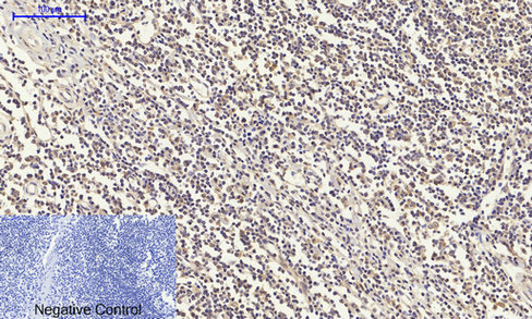
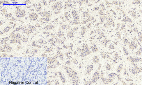
Immunohistochemical analysis of paraffin-embedded Human-stomach-cancer tissue. 1,IGF-IR Polyclonal Antibody was diluted at 1:200(4°C,overnight). 2, Sodium citrate pH 6.0 was used for antibody retrieval(>98°C,20min). 3,Secondary antibody was diluted at 1:200(room tempeRature, 30min). Negative control was used by secondary antibody only.
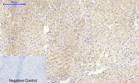
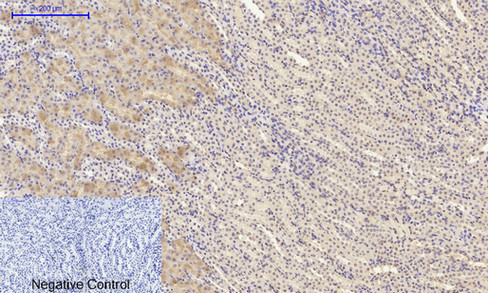
Immunohistochemical analysis of paraffin-embedded Human-liver tissue. 1,IGF-IR Polyclonal Antibody was diluted at 1:200(4°C,overnight). 2, Sodium citrate pH 6.0 was used for antibody retrieval(>98°C,20min). 3,Secondary antibody was diluted at 1:200(room tempeRature, 30min). Negative control was used by secondary antibody only.
Immunohistochemical analysis of paraffin-embedded Rat-kidney tissue. 1,IGF-IR Polyclonal Antibody was diluted at 1:200(4°C,overnight). 2, Sodium citrate pH 6.0 was used for antibody retrieval(>98°C,20min). 3,Secondary antibody was diluted at 1:200(room tempeRature, 30min). Negative control was used by secondary antibody only.
Immunohistochemical analysis of paraffin-embedded Human-Tonsil tissue. 1,IGF-IR Polyclonal Antibody was diluted at 1:200(4°C,overnight). 2, Sodium citrate pH 6.0 was used for antibody retrieval(>98°C,20min). 3,Secondary antibody was diluted at 1:200(room tempeRature, 30min). Negative control was used by secondary antibody only.
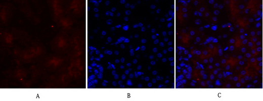
Immunofluorescence analysis of mouse-lung tissue. 1,IGF-IR Polyclonal Antibody(red) was diluted at 1:200(4°C,overnight). 2, Cy3 labled Secondary antibody was diluted at 1:300(room temperature, 50min).3, Picture B: DAPI(blue) 10min. Picture A:Target. Picture B: DAPI. Picture C: merge of A+B
Western Blot analysis of various cells using IGF-IR Polyclonal Antibody diluted at 1:2000
Immunohistochemical analysis of paraffin-embedded Mouse-kidney tissue. 1,IGF-IR Polyclonal Antibody was diluted at 1:200(4°C,overnight). 2, Sodium citrate pH 6.0 was used for antibody retrieval(>98°C,20min). 3,Secondary antibody was diluted at 1:200(room tempeRature, 30min). Negative control was used by secondary antibody only.
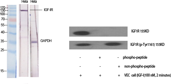
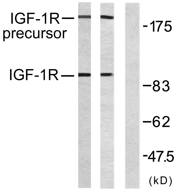
Western blot analysis of lysates from 293 cells, treated with Insulin, using IGF1R Antibody. The lane on the right is blocked with the synthesized peptide.
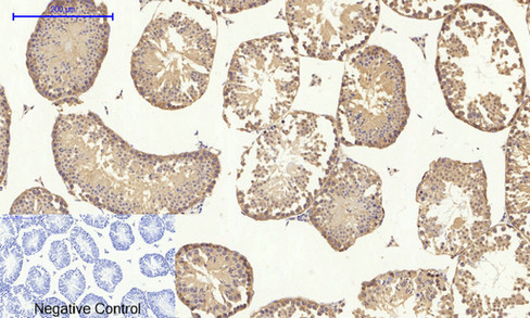
Immunohistochemical analysis of paraffin-embedded Mouse-testis tissue. 1,IGF-IR Polyclonal Antibody was diluted at 1:200(4°C,overnight). 2, Sodium citrate pH 6.0 was used for antibody retrieval(>98°C,20min). 3,Secondary antibody was diluted at 1:200(room tempeRature, 30min). Negative control was used by secondary antibody only.
Immunofluorescence analysis of mouse-lung tissue. 1,IGF-IR Polyclonal Antibody(red) was diluted at 1:200(4°C,overnight). 2, Cy3 labled Secondary antibody was diluted at 1:300(room temperature, 50min).3, Picture B: DAPI(blue) 10min. Picture A:Target. Picture B: DAPI. Picture C: merge of A+B
Immunofluorescence analysis of mouse-kidney tissue. 1,IGF-IR Polyclonal Antibody(red) was diluted at 1:200(4°C,overnight). 2, Cy3 labled Secondary antibody was diluted at 1:300(room temperature, 50min).3, Picture B: DAPI(blue) 10min. Picture A:Target. Picture B: DAPI. Picture C: merge of A+B
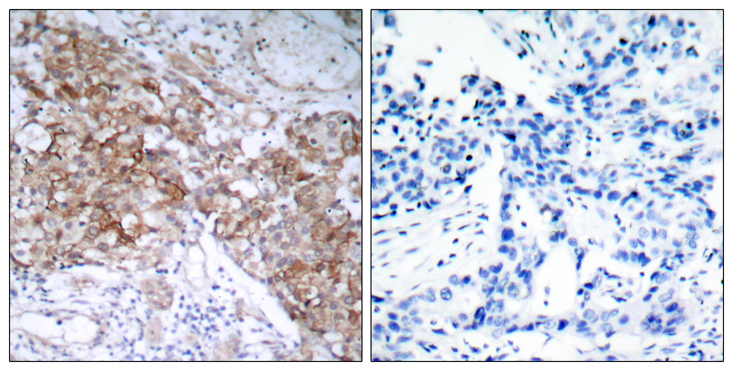
Immunohistochemistry analysis of paraffin-embedded human breast carcinoma tissue, using IGF1R Antibody. The picture on the right is blocked with the synthesized peptide.
Immunofluorescence analysis of rat-kidney tissue. 1,IGF-IR Polyclonal Antibody(red) was diluted at 1:200(4°C,overnight). 2, Cy3 labled Secondary antibody was diluted at 1:300(room temperature, 50min).3, Picture B: DAPI(blue) 10min. Picture A:Target. Picture B: DAPI. Picture C: merge of A+B
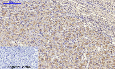
Immunohistochemical analysis of paraffin-embedded Human-liver-cancer tissue. 1,IGF-IR Polyclonal Antibody was diluted at 1:200(4°C,overnight). 2, Sodium citrate pH 6.0 was used for antibody retrieval(>98°C,20min). 3,Secondary antibody was diluted at 1:200(room tempeRature, 30min). Negative control was used by secondary antibody only.
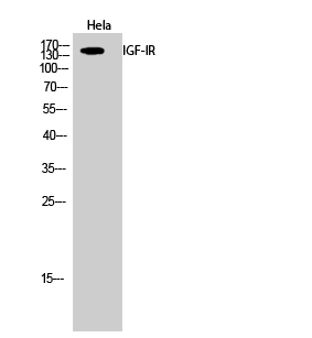
Western Blot analysis of Hela cells using IGF-IR Polyclonal Antibody diluted at 1:2000
Immunofluorescence analysis of mouse-kidney tissue. 1,IGF-IR Polyclonal Antibody(red) was diluted at 1:200(4°C,overnight). 2, Cy3 labled Secondary antibody was diluted at 1:300(room temperature, 50min).3, Picture B: DAPI(blue) 10min. Picture A:Target. Picture B: DAPI. Picture C: merge of A+B