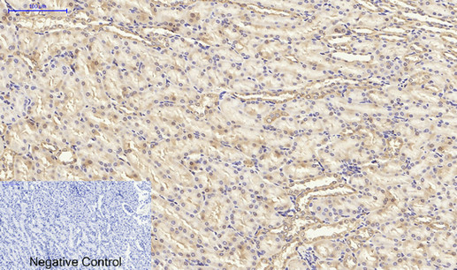
Immunohistochemical analysis of paraffin-embedded Rat-kidney tissue. 1,PPAR-γ Polyclonal Antibody was diluted at 1:200(4°C,overnight). 2, Sodium citrate pH 6.0 was used for antibody retrieval(>98°C,20min). 3,Secondary antibody was diluted at 1:200(room tempeRature, 30min). Negative control was used by secondary antibody only.
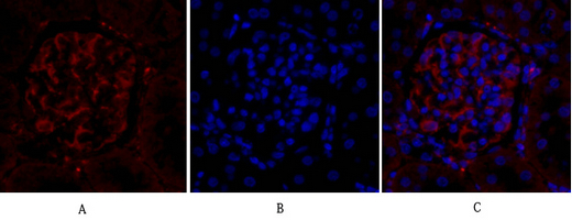
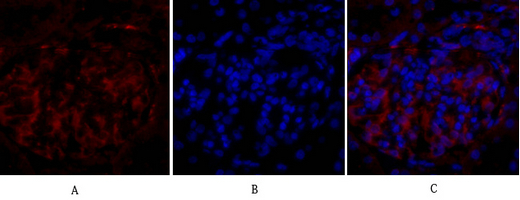
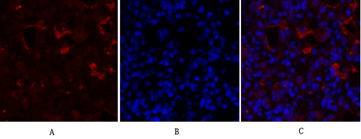
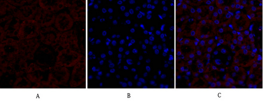
Immunofluorescence analysis of rat-kidney tissue. 1,PPAR-γ Polyclonal Antibody(red) was diluted at 1:200(4°C,overnight). 2, Cy3 labled Secondary antibody was diluted at 1:300(room temperature, 50min).3, Picture B: DAPI(blue) 10min. Picture A:Target. Picture B: DAPI. Picture C: merge of A+B
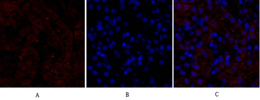
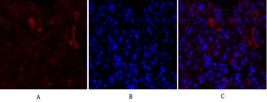
Immunofluorescence analysis of mouse-kidney tissue. 1,PPAR-γ Polyclonal Antibody(red) was diluted at 1:200(4°C,overnight). 2, Cy3 labled Secondary antibody was diluted at 1:300(room temperature, 50min).3, Picture B: DAPI(blue) 10min. Picture A:Target. Picture B: DAPI. Picture C: merge of A+B
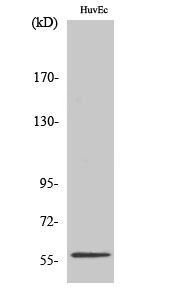
Western Blot analysis of HuvEc cells using PPAR-γ Polyclonal Antibody diluted at 1:1000
Immunohistochemical analysis of paraffin-embedded Human-Tonsil tissue. 1,PPAR-γ Polyclonal Antibody was diluted at 1:200(4°C,overnight). 2, Sodium citrate pH 6.0 was used for antibody retrieval(>98°C,20min). 3,Secondary antibody was diluted at 1:200(room tempeRature, 30min). Negative control was used by secondary antibody only.
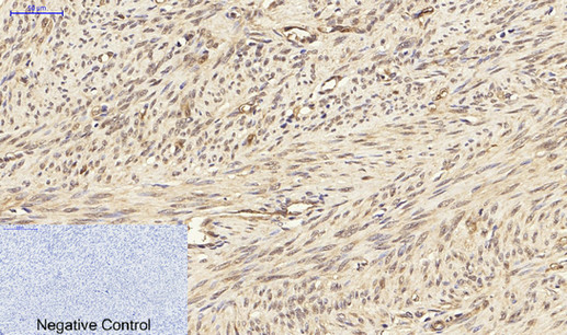
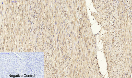
Immunohistochemical analysis of paraffin-embedded Human-uterus tissue. 1,PPAR-γ Polyclonal Antibody was diluted at 1:200(4°C,overnight). 2, Sodium citrate pH 6.0 was used for antibody retrieval(>98°C,20min). 3,Secondary antibody was diluted at 1:200(room tempeRature, 30min). Negative control was used by secondary antibody only.
Immunofluorescence analysis of rat-kidney tissue. 1,PPAR-γ Polyclonal Antibody(red) was diluted at 1:200(4°C,overnight). 2, Cy3 labled Secondary antibody was diluted at 1:300(room temperature, 50min).3, Picture B: DAPI(blue) 10min. Picture A:Target. Picture B: DAPI. Picture C: merge of A+B
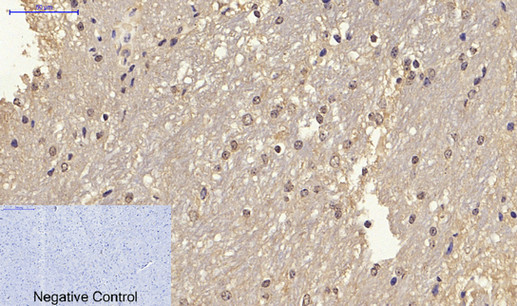
Immunohistochemical analysis of paraffin-embedded Mouse-brain tissue. 1,PPAR-γ Polyclonal Antibody was diluted at 1:200(4°C,overnight). 2, Sodium citrate pH 6.0 was used for antibody retrieval(>98°C,20min). 3,Secondary antibody was diluted at 1:200(room tempeRature, 30min). Negative control was used by secondary antibody only.
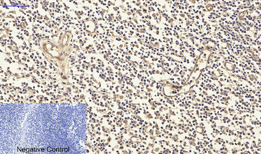
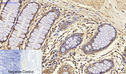
Immunohistochemical analysis of paraffin-embedded Human-colon tissue. 1,PPAR-γ Polyclonal Antibody was diluted at 1:200(4°C,overnight). 2, Sodium citrate pH 6.0 was used for antibody retrieval(>98°C,20min). 3,Secondary antibody was diluted at 1:200(room tempeRature, 30min). Negative control was used by secondary antibody only.
Immunofluorescence analysis of rat-lung tissue. 1,PPAR-γ Polyclonal Antibody(red) was diluted at 1:200(4°C,overnight). 2, Cy3 labled Secondary antibody was diluted at 1:300(room temperature, 50min).3, Picture B: DAPI(blue) 10min. Picture A:Target. Picture B: DAPI. Picture C: merge of A+B
Immunofluorescence analysis of rat-lung tissue. 1,PPAR-γ Polyclonal Antibody(red) was diluted at 1:200(4°C,overnight). 2, Cy3 labled Secondary antibody was diluted at 1:300(room temperature, 50min).3, Picture B: DAPI(blue) 10min. Picture A:Target. Picture B: DAPI. Picture C: merge of A+B
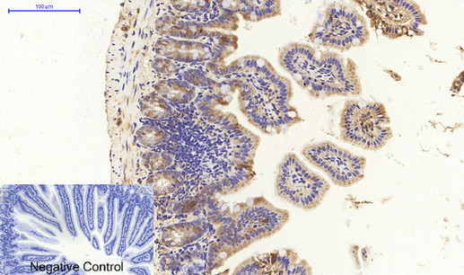
Immunohistochemical analysis of paraffin-embedded Mouse-colon tissue. 1,PPAR-γ Polyclonal Antibody was diluted at 1:200(4°C,overnight). 2, Sodium citrate pH 6.0 was used for antibody retrieval(>98°C,20min). 3,Secondary antibody was diluted at 1:200(room tempeRature, 30min). Negative control was used by secondary antibody only.
Western Blot analysis of various cells using PPAR-γ Polyclonal Antibody diluted at 1:1000
Immunofluorescence analysis of mouse-kidney tissue. 1,PPAR-γ Polyclonal Antibody(red) was diluted at 1:200(4°C,overnight). 2, Cy3 labled Secondary antibody was diluted at 1:300(room temperature, 50min).3, Picture B: DAPI(blue) 10min. Picture A:Target. Picture B: DAPI. Picture C: merge of A+B
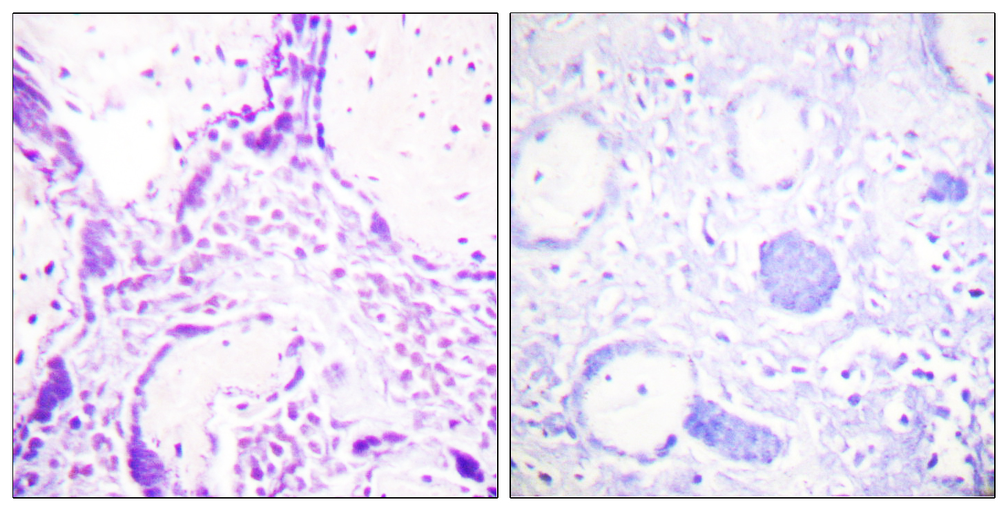
Immunohistochemistry analysis of paraffin-embedded human placenta tissue, using PPAR-gamma Antibody. The picture on the right is blocked with the synthesized peptide.
Immunohistochemical analysis of paraffin-embedded Human-uterus-cancer tissue. 1,PPAR-γ Polyclonal Antibody was diluted at 1:200(4°C,overnight). 2, Sodium citrate pH 6.0 was used for antibody retrieval(>98°C,20min). 3,Secondary antibody was diluted at 1:200(room tempeRature, 30min). Negative control was used by secondary antibody only.
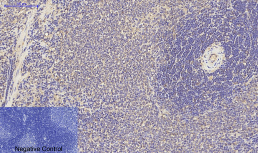
Immunohistochemical analysis of paraffin-embedded Rat-spleen tissue. 1,PPAR-γ Polyclonal Antibody was diluted at 1:200(4°C,overnight). 2, Sodium citrate pH 6.0 was used for antibody retrieval(>98°C,20min). 3,Secondary antibody was diluted at 1:200(room tempeRature, 30min). Negative control was used by secondary antibody only.
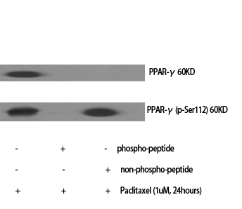
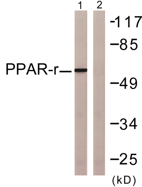
Western blot analysis of lysates from HUVEC cells, using PPAR-gamma Antibody. The lane on the right is blocked with the synthesized peptide.