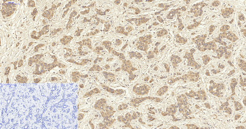
Immunohistochemical analysis of paraffin-embedded human-liver-cancer tissue. 1,VIM Polyclonal Antibody was diluted at 1:200(4° overnight). 2, Sodium citrate pH 6.0 was used for antigen retrieval(>98°C,20min). 3,Secondary antibody was diluted at 1:200(room temperature, 30min) Negtive control was used by secondary antibody only.)
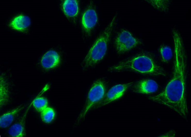
Immunofluorescence analysis of Hela cell. 1,VIM Polyclonal Antibody(green) was diluted at 1:200(4° overnight). 2, Goat Anti Rabbit Alexa Fluor 488 was diluted at 1:1000(room temperature, 50min). 3 DAPI(blue) 10min.
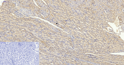
Immunohistochemical analysis of paraffin-embedded RAT-KIDNEY tissue. 1,VIM Polyclonal Antibody was diluted at 1:200(4° overnight). 2, Sodium citrate pH 6.0 was used for antigen retrieval(>98°C,20min). 3,Secondary antibody was diluted at 1:200(room temperature, 30min) Negtive control was used by secondary antibody only.)
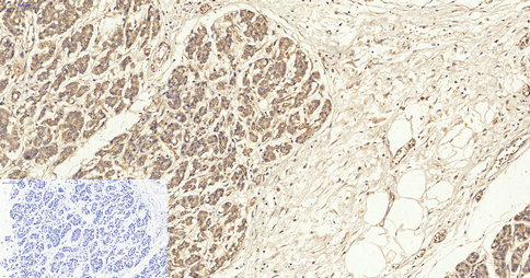
Immunohistochemical analysis of paraffin-embedded human-stomach-cancer tissue. 1,VIM Polyclonal Antibody was diluted at 1:200(4° overnight). 2, Sodium citrate pH 6.0 was used for antigen retrieval(>98°C,20min). 3,Secondary antibody was diluted at 1:200(room temperature, 30min) Negtive control was used by secondary antibody only.)
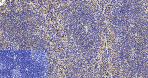
Immunohistochemical analysis of paraffin-embedded mouse-spleen tissue. 1,VIM Polyclonal Antibody was diluted at 1:200(4° overnight). 2, Sodium citrate pH 6.0 was used for antigen retrieval(>98°C,20min). 3,Secondary antibody was diluted at 1:200(room temperature, 30min) Negtive control was used by secondary antibody only.)
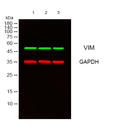
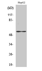
Western Blot analysis of HepG2 cells using VIM Polyclonal Antibody diluted at 1:1000. Secondary antibody was diluted at 1:20000
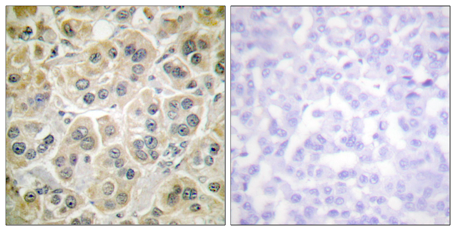
Immunohistochemistry analysis of paraffin-embedded human breast carcinoma tissue, using Vimentin Antibody. The picture on the right is blocked with the synthesized peptide.
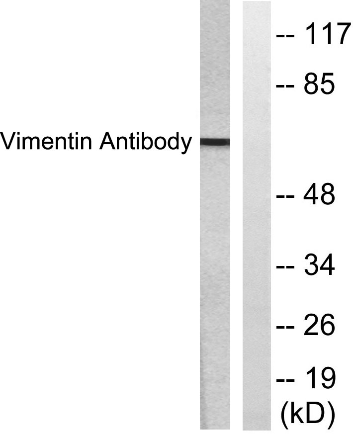
Western blot analysis of lysates from HepG2 cells, treated with Adriamycin 0.5uM 5h, using Vimentin Antibody. The lane on the right is blocked with the synthesized peptide.
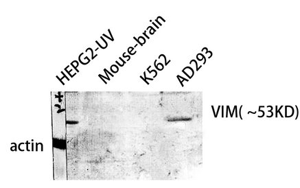
Western Blot analysis of HepG2-UV MOUSE-BRAIN AD293 K562 cells using VIM Polyclonal Antibody diluted at 1:1000. Secondary antibody was diluted at 1:20000