Product overview

Full product name
Code
Host
Isotype
Size
Clonality
Immunogen
Purification
Concentration
Formulation
Storage
Background
MAP1A and MAP1B are microtubule-associated proteins which mediate the physical interactions between microtubules and components of the cytoskeleton. MAP1A and MAP1B each consist of a heavy chain subunit and multiple light chain subunits. The protein encoded by this gene is one of the light chain subunits and can associate with either MAP1A or MAP1B. Two transcript variants encoding different isoforms have been found for this gene. The expression of variant 1 is suppressed in many tumor cell lines, suggesting that may be involved in carcinogenesis.
Uniprot accession
Molecular weight
Gene ID
Synonyms
Research area
Target protein
Recommended dilution
| Application | Dilution |
|---|---|
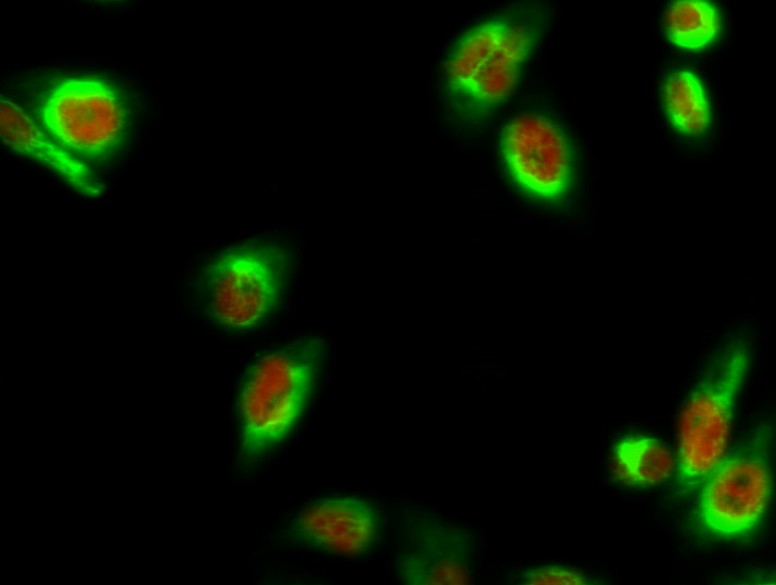
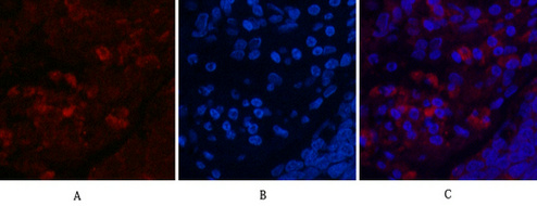
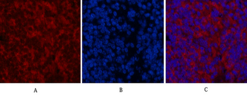
| IF | 1:200 |
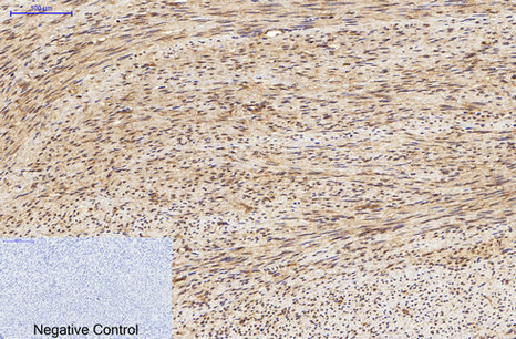
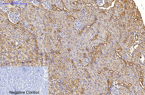
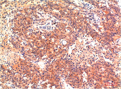
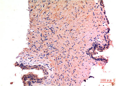
| IHC | 1:100-200 |
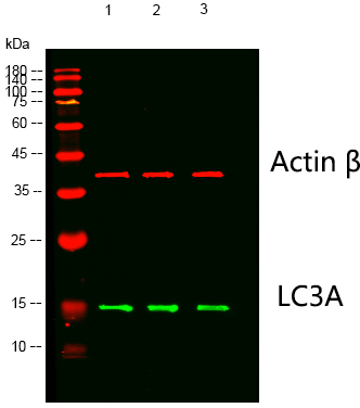
| WB | 1:1000-2000 |
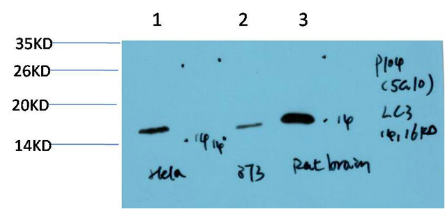
Images
Protocols
Customer reviews and Q&As