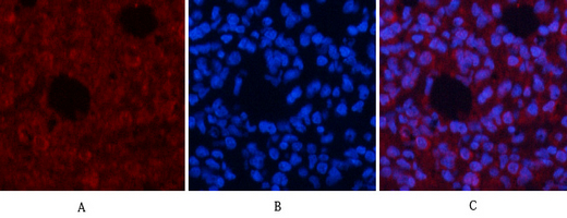
Immunofluorescence analysis of human-lung tissue. 1,AP-1 Polyclonal Antibody(Red) was diluted at 1:200(4°C overnight). 2, Cy3 labled Secondary antibody was diluted at 1:300(room temperature, 50min).3, Picture B: DAPI(blue) 10min. Picture A:Target. Picture B: DAPI. Picture C: merge of A+B
Immunofluorescence analysis of human-lung tissue. 1,AP-1 Polyclonal Antibody(Red) was diluted at 1:200(4°C overnight). 2, Cy3 labled Secondary antibody was diluted at 1:300(room temperature, 50min).3, Picture B: DAPI(blue) 10min. Picture A:Target. Picture B: DAPI. Picture C: merge of A+B
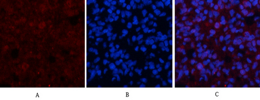
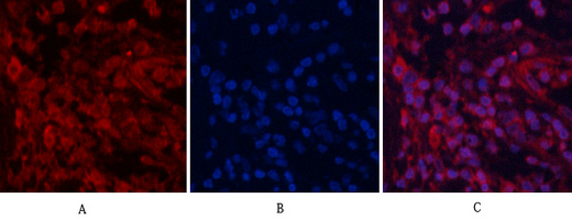
Immunofluorescence analysis of human-stomach tissue. 1,AP-1 Polyclonal Antibody(Red) was diluted at 1:200(4°C overnight). 2, Cy3 labled Secondary antibody was diluted at 1:300(room temperature, 50min).3, Picture B: DAPI(blue) 10min. Picture A:Target. Picture B: DAPI. Picture C: merge of A+B
Immunofluorescence analysis of human-stomach tissue. 1,AP-1 Polyclonal Antibody(Red) was diluted at 1:200(4°C overnight). 2, Cy3 labled Secondary antibody was diluted at 1:300(room temperature, 50min).3, Picture B: DAPI(blue) 10min. Picture A:Target. Picture B: DAPI. Picture C: merge of A+B
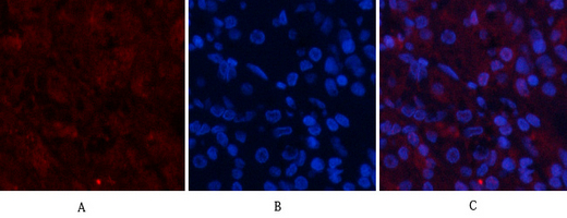
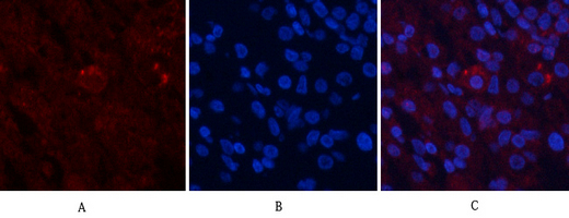
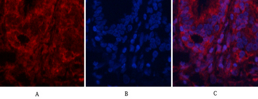
Immunofluorescence analysis of rat-lung tissue. 1,AP-1 Polyclonal Antibody(Red) was diluted at 1:200(4°C overnight). 2, Cy3 labled Secondary antibody was diluted at 1:300(room temperature, 50min).3, Picture B: DAPI(blue) 10min. Picture A:Target. Picture B: DAPI. Picture C: merge of A+B
Immunofluorescence analysis of rat-lung tissue. 1,AP-1 Polyclonal Antibody(Red) was diluted at 1:200(4°C overnight). 2, Cy3 labled Secondary antibody was diluted at 1:300(room temperature, 50min).3, Picture B: DAPI(blue) 10min. Picture A:Target. Picture B: DAPI. Picture C: merge of A+B
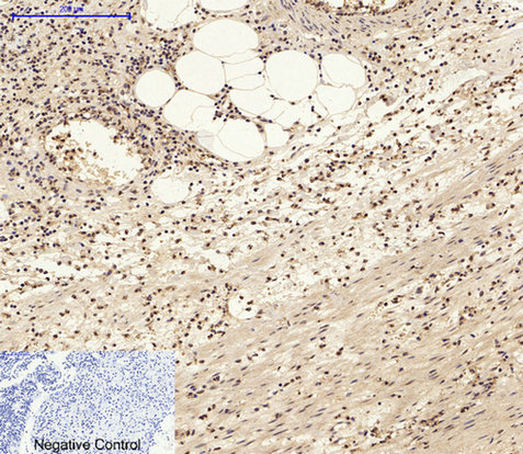
Immunohistochemical analysis of paraffin-embedded Human uterus. 1, Antibody was diluted at 1:100(4°C overnight). 2, High-pressure and temperature EDTA, pH8.0 was used for antigen retrieval. 3,Secondary antibody was diluted at 1:200(room temperature, 30min).
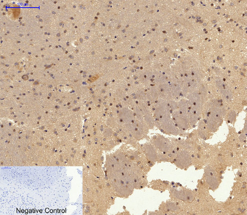
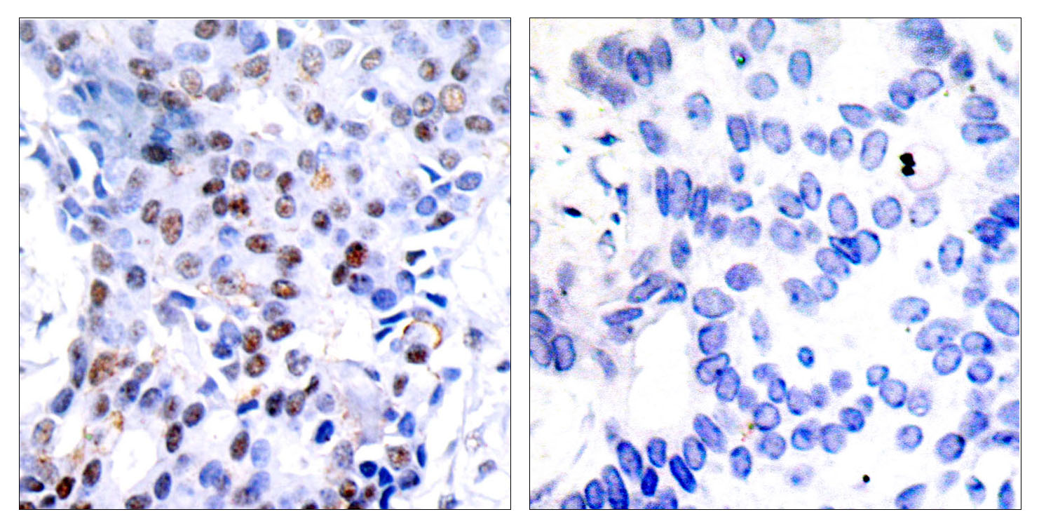
Immunohistochemical analysis of paraffin-embedded Rat-brain tissue. 1,AP-1 Polyclonal Antibody was diluted at 1:200(4°C overnight). 2, Sodium citrate pH 6.0 was used for antibody retrieval(>98°C,20min). 3,Secondary antibody was diluted at 1:200(room temperature, 30min). Negative control was used by secondary antibody only.
Immunohistochemistry analysis of paraffin-embedded human breast carcinoma tissue, using c-Jun Antibody. The picture on the right is blocked with the synthesized peptide.
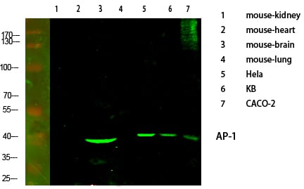
Western Blot analysis of various cells using primary antibody diluted at 1:1000(4°C overnight). Secondary antibody:Goat Anti-rabbit IgG IRDye 800( diluted at 1:5000, 25°C, 1 hour).
Western Blot analysis of various cells using AP-1 Polyclonal Antibody diluted at 1:2000
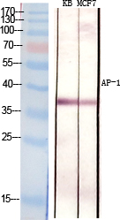
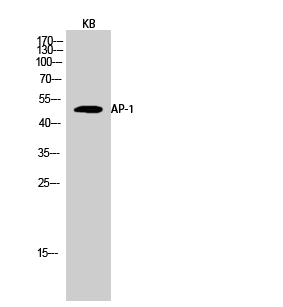
Western Blot analysis of KB cells using AP-1 Polyclonal Antibody diluted at 1:2000
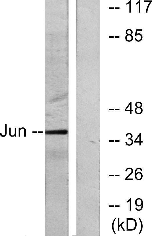
Western blot analysis of lysates from HeLa cells, using c-Jun Antibody. The lane on the right is blocked with the synthesized peptide.